


Native chromatin immunoprecipitation protocol
实验概要
The method is a native chromatin immunoprecipitation protocol.
主要试剂
1. 10 x TBS
0.1 M Tris-HCl (pH 7.5)
1.5 M NaCl
30 mM CaCl2
20 mM MgCl2
50 mM Na butyrate (pH 8.0)
2. Digestion buffer
0.32 M sucrose
50 mM Tris-HCl (pH 7.5)
4 mM MgCl2
1 mM CaCl2
0.1 mM PMSF
5 mM Na butyrate
3. Lysis buffer
1.0 mM Tris-HCl (pH7.4)
0.2 mM Na2EDTA 0.2 mM PMSF
5 mM Na butyrate
4. Incubation buffer
50 mM NaCl
20 mM Tris-HCL (pH 7.5)
20 mM Na butyrate
5 mM Na2EDTA
0.1 mM PMSF
5. Buffer A
50 mM Tris-HCl, (pH 7.5)
10 mM EDTA
5 mM Na butyrate
50 mM NaCl
6. Buffer B
50 mM Tris-HCL (pH 7.5)
10 mM EDTA
5 mM Na butyrate
100 mM NaCl
7. Buffer C
50 mM Tris-HCL (pH 7.5)
10 mM EDTA
5 mM Na butyrate
150 mM NaCl
8. Protein A Sepharose
Pre-swell protein A Sepharose overnight in buffer A at 4°C. Centrifuge (10,000 x g, 10 min) and resuspend pellet in approximately an equal volume (50% v/v) of buffer A.
实验步骤
1. The preparation of native chromatin from cultured human cells
1) Cultured cells (e.g. HL-60 or lymphoblastoids) are grown to a density of approximately 1 x 10⁶ cells/ml, until they are in log phase.
2) Harvest cells: centrifuge samples (7,000 g, 10 min, 4°C) and wash the cell pellet 3 x ice cold PBS (Phosphate buffered saline).
It is essential that 5 mM Na butyrate is present in all solutions throughout chromatin isolation when using antibodies to acetylated histones to prevent deacetylation. |
3) Resuspend cell pellet in TBS (Tris buffered saline) at 2 x 10⁷ cells/ml and add an equal volume of 1.0% v/v Tween 40 in TBS. Add PMSF to a final concentration of 0.5 mM. Leave stirring gently on ice for 1 hr (Transfer the suspension into a 50 ml tube with a small magnetic bar or flea; place the tube in ice on top of a magnetic stirrer).
4) Transfer cell lysate to an all-glass homogenizer and homogenize 7 ml aliquots with seven strokes using an ‘A’ or ‘tight’ pestle. Check that nuclei have been released by phase-contrast microscopy; intact cells should have the central dark region of the nucleus surrounded by a halo, which is the less dense cytoplasm.
You may have to increase or decrease this homogenization step to maximize the yield of nuclei depending on cell line. |
5) Centrifuge samples (10,000 g, 20 min, 4°C).
6) Resuspend nuclei pellet in 25% [w/v] sucrose/TBS at 4x10⁶ nuclei / ml and underlay with 0.5 vol of 50% [w/v] sucrose / TBS; centrifuge the samples (14,000 g, 25 min, 4°C).
7) Discard supernatant and wash nuclei pellet in 5 ml 25% [w/v] sucrose/TBS; centrifuge samples (14,000 x g, 25 min, 4°C).
8) Resuspend nuclei pellet in 5 ml digestion buffer and check absorbance ratios at 260 nm and 280 nm for a diluted sample of the nuclei suspension; calculate the approximate DNA concentration from the A260 reading (the ratio of A260/A280 should be about 1.1). Centrifuge samples (10,000 rpm, 10 min, 4°C) and resuspend the nuclei pellet at 0.5 mg/ml in 1.7 ml Eppendorf tube(s).
2. Micrococcal nuclease digestion
Normally we add 50 U micrococcal nuclease per 0.5 mg DNA, in a reaction volume of 1.0 ml. This is usually provided as a powder; dissolve the micrococcal nuclease in dH 20 to the required concentration and store as small aliquots at -20°C. Aliquots may be re-frozen and re-used once. This step needs to be carefully controlled, especially in the initial preparations.
High concentrations of micrococcal nuclease may over-digest the chromatin, leading to sub-nucleosomal particles. You should aim to obtain a long/medium oligonucleosome ladder. If pure mononucleosome preparations are required carry out a linear sucrose gradient (5-20%), this will increase resolution. |
1) Perform microccal nuclease digestions at 37°C for 5 min.
2) Stop reaction by addition of 0.2 M EDTA to a final concentration of 5 mM.
3) Place all samples on ice for 5 min; centrifuge samples (8,000 g, 5 min).
4) Remove and keep the first S/N (this is called the S1 fraction; total vol 1.0 ml); store overnight at 4°C.
5) Resuspend the pellet in 1.0 ml Lysis buffer and dialyse overnight against 2 l of the same buffer.
6) After overnight dialysis centrifuge samples (500 g, 10 min, 4°C).
7) Remove and keep the supernatant (called the S2 fraction; total vol about 1.2 ml after dialysis); store at 4°C.
8) Resuspend insoluble pelleted material in 200 μl lysis buffer (called the P fraction).
3. Analysis of soluble chromatin fractions
1) Check A260/A280 in all samples; the ratios for S1, S2 and P fractions are approximately 1.7, 1.5 and 1.3 respectively. Analyze all samples by 1.2% agarose gel electrophoresis.
Do not place ethidium bromide in the agarose gel or the electrophoresis buffer, because of the presence of SDS (see below) |
2) Preparation of samples: x μl (total of 5 μg) chromatin fraction (S1, S2 and P) y μl dH2O (x y = 25 μl) 3 μl 1% [w/v] SDS (final conc 0.1%) 2 μl gel loading buffer, containing bromophenol blue
3) Stain the gel with 0.5 μg/ml ethidium bromide after the run has finished.
4. Immunoprecipitation
1) 100-200 μg unfixed chromatin 100-200 μl affinity purified antibody (50-100 μg Ig) and the final volume made up to 1.0 ml with incubation buffer. A negative control, with no added antibody, also needs to be set up to test for any nonspecific binding of the chromatin to the protein A Sepharose.
2) Incubate overnight at 4°C on a slow rotating turntable. Add 200 μl 50% v/v protein A Sepharose; use a siliconized pipette with the tip cut off to make this step easier. Incubate for 3 h at room temperature on a fast rotating turntable. (Make sure that the Sepharose is in a suspension at all times).
3) Centrifuge samples (3,000 g, 10 min, 4°C), remove and keep the S/N; this is the unbound (or “U”) fraction.
4) Resuspend the Sepharose pellet in 1 ml buffer A and layer onto 9 ml of the same buffer using a siliconised pasteur pipette and siliconized 15 ml tube.
5) Centrifuge samples (10,000 g, 10 min, 4°C), discard the S/N and wash the Sepharose sequentially in 10 ml buffer B and buffer C.
6) Finally, resuspend the Sepharose in 1 ml buffer C and transfer back to siliconized Eppendorfs.
7) Centrifuge samples (3,000 g, 10 min, 4°C) and resuspend the sepharose pellet in 250 μl 1.0% SDS / incubation buffer and incubate for 15 min at RT on a fast turntable. (Ensure that the Sepharose is thoroughly resuspended at all times).
8) Centrifuge the samples (3,000 g, 10 min, 4°C) and remove and keep S/N; this is the bound (or “B”) fraction.
9) Wash the sepharose in 250 μl 1.0% SDS / incubation buffer and centrifuge immediately (3,000 g, 10 min, 4°C). Remove the S/N and pool with the previous bound fraction from the previous step.
5. DNA Isolation
Add 500 μl incubation buffer to each bound fraction, to reduce the SDS concentration to 0.5% SDS.Unbound and bound fractions then treated as follows:
1) Add 0.33 vol (330 μl) phenol/chloroform; vortex and spin (13,000 rpm, 10 min, microcentrifuge). Keep the organic phase and interface; this is used to isolate immunoprecipitated proteins (see below).
2) Transfer the aqueous supernatant to an equal volume (1.0 ml) of phenol/chloroform; vortex and spin (13,000 rpm, 10 min, microcentrifuge)
3) Transfer supernatant to an equal volume (1.0 ml) of chloroform; vortex and spin (13,000 rpm, 10 min, microcentrifuge)
4) Transfer S/N to a clean centrifuge tube and add 0.1 vol (100 μl) 4 M LiCl, 50 μg glycogen (Molecular biology grade, dissolved in dH20 at 2 mg/ml) as a carrier and 4 vol of ethanol. Vortex thoroughly and leave at -20°C overnight.
5) Centrifuge samples (13,000 g, 15 min) to precipitate the DNA.
6) Wash the pellet with 70% ethanol and redissolve the DNA in 250 μl TE buffer.
7) Store samples at -20°C or proceed with detection method (PCR, microarray, etc).
8) PCR is used to quantify DNA levels of specific loci. This is analyzed semi-quantitatively (analyses of PCR endproduct by agarose gel) using primers which can be designed using this tool.
Alternatively, DNA levels are quantitatively measured by real-time PCR. Primers and probes are often designed using software provided with the real-time PCR apparatus.
6. Protein Isolation
1) To the first phenol/chloroform phase (see DNA isolation; step1) add 5 μl of a 1 mg/ml solution of BSA (to be used as a carrier), 0.01 vol (4 μl) 10 M H2SO4 and 12 vol of acetone.
2) After precipitation at -20°C wash the protein pellets once in acidified acetone (1:6 100 mM H2SO4:acetone) and 3 times in dry acetone. Proteins can be analyzed by SDS-PAGE.


-
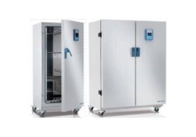
Thermo Scientific Heratherm 大容量高端型烘箱(Thermo Scientific Heratherm Large Capacity Advanced Protocol Ovens)
询底价 -
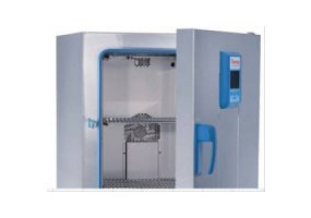
Thermo Scientific Heratherm 高端安全型烘箱(Thermo Scientific Heratherm Advanced Protocol Security Ovens)
询底价 -
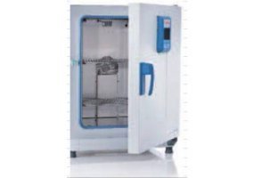
Thermo Scientific Heratherm 高端型烘箱(Thermo Scientific Heratherm Advanced Protocol Ovens)
询底价 -
Thermo Scientific Heratherm 通用型烘箱(Thermo Scientific Heratherm General Protocol Ovens)
询底价


